Optical Coherence Tomography HHO-2000 - Hans Heiss

Optical Coherence Tomography HHO-2000

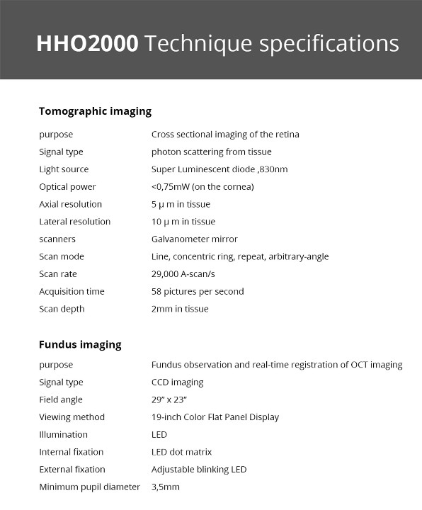
Optical coherence tomography (OCT) is a new, noninvasive, noncontact, transpupillary imaging technology which can image retinal structures in vivo with a resolution of 5 to 8 microns. Cross-sectional images of the retina are produced using the optical backscattering of light in a fashion analogous to B- scan ultrasonography and cofocal microscopy. Cross-sectional images of the retina, is revolutionizing the early detection and treatment and greatly enhanced our quality of patient care.

Applications

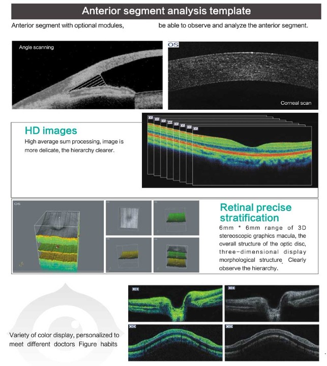
In vivo, cross-sectional images and quantitative analysis of retinal features to optimize the diagnosis and monitoring of retinal disease and for enhanced pre - and post-therapy assessment.

High-quality images and accurate measures RNFL and the optic nerve head to aid in the detection and management of glaucoma.

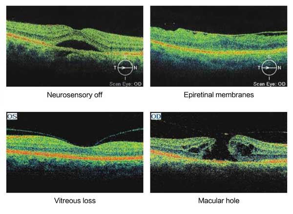
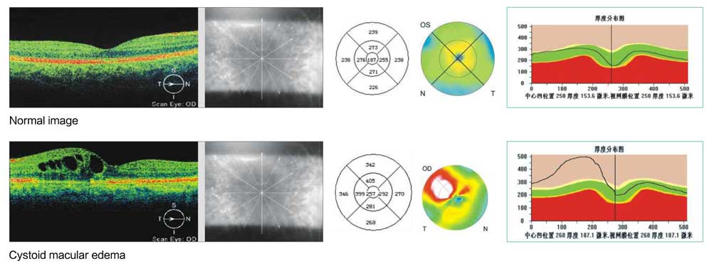
Cross-sectional images are valuable for clinical evaluation of macular holes, macular edema and other retinal pathologies.

Precise location of pathology to expand diagnostic confidence and therapeutic precision .

Normal and abnormal image contrast image

Lesions image by image with normal OCT images, the regional thickness values? topographic maps, diagrams and other multi-thickness contrast, thereby comprehensive judgment of disease.

Advantages

High performance at a low price

Modular design increases flexibility, reusability and maintainability. We can provide personalized design according to the customers' needs.

With the powerful software, has clear, easy-to-use interface and supports multi-language.


Specifications: HHO2000

SPECIFICATIONS

Tomographic imaging
purpose Cross sectional imaging of the retina
Signal type photon scattering from tissue
Light source Super Luminescent diode ,830nm
Optical power <0,75mW (on the cornea)
Axial resolution 5 μ m in tissue
Lateral resolution 10 μ m in tissue
scanners Galvanometer mirror
Scan mode Line, concentric ring, repeat, arbitrary-angle
Scan rate 29,000 A-scan/s
Acquisition time 58 pictures per second
Scan depth 2mm in tissue
Fundus imaging
purpose Fundus observation and real-time registration of OCT imaging
Signal type CCD imaging
Field angle 29” x 23”
Viewing method 19-inch Color Flat Panel Display
Illumination LED
Internal fixation LED dot matrix
External fixation Adjustable blinking LED
Minimum pupil diameter 3,5mm

Inquiry about

Accept Terms and Conditions
Optical Coherence Tomography HHO-2000

You Recently Viewed Items

Events / Shows View All

AAO 2023
AAO 2023

AAO 2023

Bascom Palmer 2023
Bascom Palmer 2023

Bascom Palmer 2023

Reunión virtual con el Dr. Julian Triviño (Colombia)

Reunión virtual con el Dr. Julian Triviño (Colombia)

AAO 2021
AAO 2021

 Friday, Nov. 12 - Monday, Nov. 15  Ernest N. Morial Convention CenterNew Orleans, LA.

5555 NW 74 AVE.
MIAMI. FL. 33166
P: 305-882-0120
F: 305-882-0140
sales@mercoframes.net
Skype mercoframesonline
Skype sales3mercoframes
Skype helpmercoframes
WhatsApp +1 305-882-0120
Hangouts ale@mercoframes.net