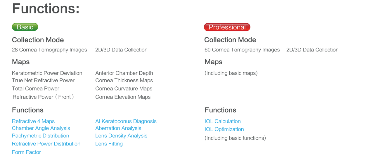
Scansys provides a professional solution for anterior segment diagnosis. It applies Scheimpflug camera which can collect 107520/230400 data points and generates 28/60 cornea tomography images in high resolution. Scansys can provide a series of topography maps including cornea curvature maps, cornea thickness maps, cornea elevation maps, etc.
◉ Keratoconus Diagnosis
Scansys can provide the prevalence of Keratoconus by using the AI algorithm.Further checking the topographic maps to accurately analyze amd diagnose the keratoconus.
◉ Refractive surgery
Total cornea aberration guides surgeons to evaluate preoperative and postoperative visual quality to ensure patients of best surgery effect.
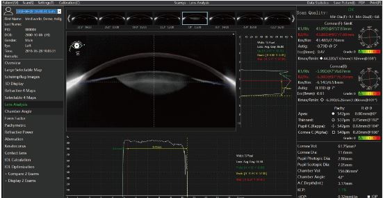
◉ IOL Optimization
Specially designed for cataract surgery. It supports clinicians to choose suitable Toric IOL、 Aspheric IOL or Multifocal IOL for patients.
◉ ICL Surgery Examination
Scansys supports in different angles to collect a high- resolution picture, It also provides White to White, AC depth for ICL surgery.
AI intelligence recommends the diameter of the ICL crystal and gives elevation of the arch.

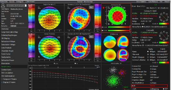
This anterior segment analyzer clinically collects a large amount of data from all over the world and introduces AI algorithms to more intelligently determine the possibility of cone disease (KCP) in the current case. (Reference value: KCP, range 0% -100%) The above figure contains the topographic map of Refractive 4 Maps, the axial curvature map of the rear surface, and the trend distribution of the thickness map. These are the key references for judging the keratoconus.

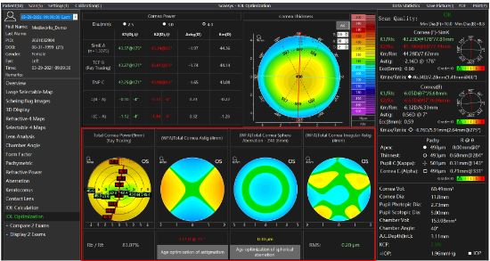
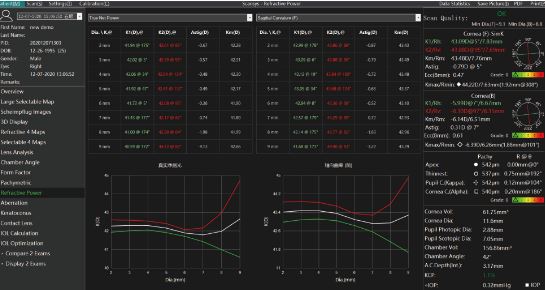
Specially designed for the “IOL Optimization”of refractive cataract surgery, The anterior segment analyzer is given the K1, K2, Km, and Astig values of the three types of corneal refractive power (Simk, total corneal power, true net refractive power), and Kappa&Alpha angle, respectively. It also provides professional data of the total corneal astigmatism aberration, total corneal spherical aberration, and the total corneal irregular astigmatism, and analysis support for solving spherical refractive errors, astigmatism, spherical aberration, and presbyopia in cataract surgery.
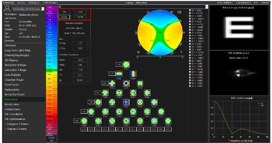
This view is a Zernike analysis of the measured corneal front,back and all surface height data, which calculates a factor for each Zernike polynomial term that describes the contribution of this polynomial to the height data.To guide the visual quality analysis of refractive surgery.

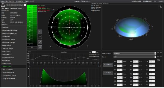
Based on the topography maps generated by Scansys,the system can recommend several lenses suitable for patinent’s cornea and simulate the images of patinent’s wearing lenses, the user can judge whether the lens is suitable. This will accelerate the work flow of lens fitting and reduce the number of trials in the actual process.

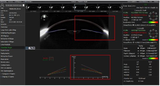
Scansys can calculate a chamber angle value based on the tomography images and its exclusive AOD graph gives a trend analysis for the distance between cornea back surface to iris. It also provides cornea volume, anterior chamber volume and anterior chamber depth calculation. These analyses is helpful to glaucoma diagnosis.

Scansys supports in any Angle to collect a high- definition picture, to provide effective data support for ICL surgery. AI intelligence recommends the diameter of the ICL crystal and gives elevation of the arch.
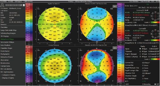
Click on the “Selectable 4 Maps” to open a window contains 4 optional color maps. Corneal thickness and elevation, etc. can be loaded into one of any 4 fields. With this option, the user can view and print out important topographic maps needed for daily work in one interface.
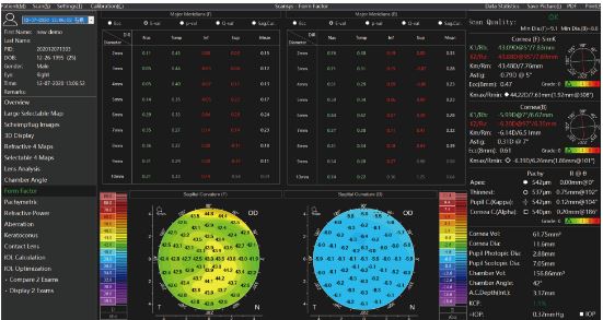
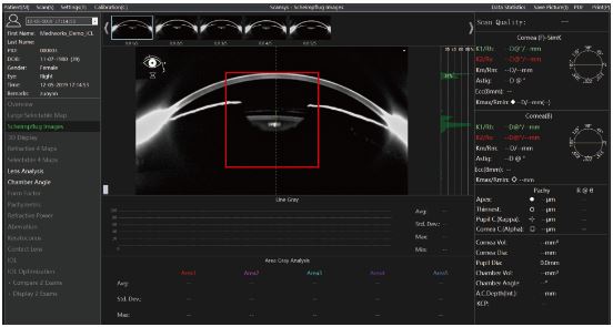
The chart upper depicts the corneal form factor and the curvature of the anterior and posterior surface of the cornea at the intersection of the radial axis and the four radial directions. Corneal shape factors include Ecc, E, Q, and P, which can be changed in the “Map and Data Settings” -> “Form Factor Presentation” in the menu bar “Settings” options
Scansys calculates the lens density value for cross section and longitudinal section which is helpful in cataract diagnosis.

In the key parameter column on the right, we give K1, K2, Km, Astig. These values are obtained in the range of 3mm in diameter of the membrane. In order to describe in more detail the difference of these values in each diameter range, We give K1, K2, Km, Astig of the axial curvature of the anterior and posterior corneal, anterior surface refractive power, true net refractive power, full corneal refractive power topographic map, the distribution table of various areas from 2mm to 9mm in diameter, and the distribution curve .The changes of these values in different topographic maps and different diameter ranges are described more intuitively and in detail.